
In the world of diagnostic imaging, visual skills can make or break a diagnosis. Imagine being able to see beyond the surface and grasp intricate details that others might miss. That’s where diag image comes into play, transforming how we perceive and understand complex medical information. With advances in technology and techniques, enhancing your visualization skills is not just beneficial; it’s essential for any practitioner in the field.
Whether you’re a seasoned radiologist or an eager student, mastering these skills opens doors to more accurate assessments and improved patient care. Let’s dive deeper into what diag image really means and explore effective strategies to elevate your visualization abilities!
What is Diag Image?
Diag image refers to the visual representation of diagnostic data, often used in medical settings. It encompasses a range of imaging techniques such as X-rays, MRIs, and CT scans.
These images play a crucial role in identifying health issues. They help healthcare professionals visualize internal structures and detect abnormalities.
With advancements in technology, diag images have become more detailed and accessible. This enhances the accuracy of diagnoses and treatment plans.
Moreover, diag images are not limited to medicine; they find applications in engineering, architecture, and even art. The concept emphasizes clarity in communicating complex information through visuals.
In essence, diag image serves as a bridge between raw data and meaningful insights across various fields. Understanding its nuances can significantly impact decision-making processes.
Understanding the Importance of Visualization in Diag Image
Visualization plays a crucial role in diag image. It involves creating mental images that help professionals interpret complex data effectively.
Accurate diagnosis often hinges on the ability to visualize what’s happening within the body. This skill allows radiologists and healthcare providers to identify abnormalities, assess conditions, and develop treatment plans with precision.
Moreover, visualization fosters communication among team members. When everyone shares a clear mental picture of findings, collaboration improves significantly. Discussions become more informed and targeted.
The importance of visualization extends beyond individual cases. In research and education, it aids in teaching anatomical structures or understanding pathological processes.
Thus, enhancing visualization skills not only benefits practitioners but also ultimately contributes to better patient outcomes. This makes it an essential aspect of diagnostic imaging practice today.
The Science Behind Visualization
Visualization taps into the brain’s remarkable ability to create mental images. This process engages various neural pathways, enhancing cognitive function and spatial awareness.
When we visualize, our brains simulate experiences as if they are real. This activation can strengthen memory retention and improve understanding of complex concepts. For those in diagnostic imaging, this skill becomes crucial.
Studies show that effective visualization can lead to quicker decision-making. It allows practitioners to interpret diag images with greater accuracy and detail.
Moreover, visualization techniques promote creativity in problem-solving; they invite innovative approaches when analyzing anatomy or pathology. The scope of what you see expands beyond mere observation it transforms how you connect the dots between data points in an image.
Techniques for Improving Visualization Skills
Improving visualization skills requires practice and a variety of techniques. One effective method is to engage in mental imagery exercises. Start by closing your eyes and picturing anatomical structures or complex images related to diag image.
Another approach is drawing what you visualize. Sketching can help solidify your understanding of spatial relationships within the body, enhancing memory retention.
Utilizing 3D modeling software also offers an interactive way to explore anatomy from different angles. This hands-on experience deepens comprehension far beyond traditional methods.
Additionally, practicing with real-life cases helps reinforce visual acuity. Analyzing actual diagnostic images fosters a more profound connection between theory and application.
Collaborating with peers can provide fresh perspectives on challenging subjects, allowing for shared experiences that enhance learning outcomes together.
Visualizing Anatomy and Pathology
Visualizing anatomy and pathology is a crucial skill in the field of diag image. It allows practitioners to identify abnormalities effectively and make informed decisions.
Anatomy visualization provides a roadmap of the body’s structures. Understanding spatial relationships between organs enhances diagnostic accuracy.
Pathology, on the other hand, tells a story about disease processes. By visualizing tissue changes, one can detect early signs of conditions that might otherwise go unnoticed.
Using advanced imaging techniques like MRI or CT scans elevates these skills further. These technologies render complex anatomical details with clarity, helping specialists pinpoint issues quickly.
Engaging with 3D models can also be beneficial. They allow for interactive learning and foster deeper comprehension of complicated systems within the human body.
A well-developed ability to visualize both normal anatomy and pathological findings enriches clinical practice and improves patient outcomes significantly.
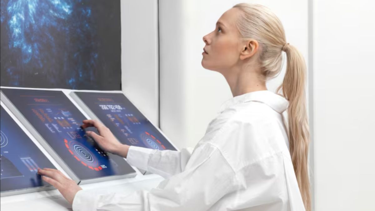
Utilizing Technology for Enhanced Visualization
Technology plays a crucial role in enhancing visualization skills within diag image practices. Modern imaging software and advanced scanning techniques provide clearer, more detailed images than ever before.
Artificial intelligence is revolutionizing the field by offering predictive analytics and pattern recognition capabilities. This allows practitioners to identify potential issues faster and with greater accuracy.
3D modeling tools transform traditional 2D images into immersive visual representations. These models help clinicians visualize complex structures, making it easier to plan interventions or understand intricate anatomy.
Virtual reality applications are gaining traction as well. They allow professionals to interact with diagnostic images in real time, creating an engaging learning experience that sharpens visualization skills.
Incorporating these technological advancements not only improves understanding but can also enhance overall patient care by ensuring accurate diagnoses and effective treatment plans.
Incorporating Visualization into Diagnostic Imaging Practice
Incorporating visualization into diagnostic imaging practice is essential for accurate interpretation. It allows radiologists to see beyond the images, enhancing their understanding of complex cases.
Start by integrating 3D modeling software. This technology offers a new perspective on traditional scans. It helps in visualizing intricate anatomical structures and relationships.
Regular training sessions can also foster better visualization skills among staff. Encourage team discussions around challenging cases to promote collaborative learning.
Utilize case studies that highlight specific pathologies and corresponding imaging findings. This method reinforces knowledge while stimulating critical thinking.
Moreover, consider engaging with virtual reality tools designed for medical education. They provide immersive experiences that enhance spatial awareness and cognitive recall.
By adopting these approaches, healthcare professionals can improve diagnostic accuracy and patient outcomes significantly through enhanced visualization techniques.
Conclusion
Understanding and enhancing your visualization skills is vital in the realm of diag image. Visualization not only aids in interpreting complex information but also empowers professionals to make informed decisions quickly. By embracing various techniques, from mental imagery practices to leveraging cutting-edge technology, you can refine your ability to visualize anatomy and pathology effectively.
Integrating these methods into your daily diagnostic imaging routine fosters a more comprehensive understanding of intricate cases. As you develop this skill set, you’ll likely notice improvements not just in accuracy but also in efficiency. The journey toward mastering visualization may take time, but the benefits it brings to diagnostics are undeniable. Embrace these strategies and watch as they transform your approach to diag image work.